23. Ultrasound
- We live in a sea of electro-magnetic waves which were generated according to a theory which says that the birth of the universe commenced with a big bang. In fact a vast majority of the events within our body involve electrical activity.
- Sound waves belong to a different genre. While the production of sound or words are a result of thoughts involving several systems which use chemical or electrical systems the actual basis of sound is a mechanical phenomena.
- Air is a compressible, elastic medium and the exhalation of air, as while speaking tends to squeeze this medium which when it re-expands sends forth a wave like pattern of compression and expansion producing a sound wave.
- In the ear the process is reversed where the initial apparatus for reception is mechanical in nature (the drums and the osscicles) but this movement is later perceived as sound by way of electro-chemical activity through the auditory nerves in the brain.
- Sound waves travel at different speeds depending on the medium and have different wavelengths. This speed is described in cycles per second. For example, sound will travel slightly faster if the atmospheric medium is hotter and travels almost three times its normal speed in water.
- The shorter the wave length and the higher the cycles per second (frequency) the more linear is its propagation. A sound like ‘aah’ has a longer wavelength and tends to spread. A whistle or a siren is of a shorter wavelength and has a greater frequency and tends to follow a more linear course. The frequency is measured by a unit called Hertz.
- A sound when it confronts a solid object echoes and returns in the direction from which it originated for example, echoing valleys or whispering galleries. The clarity of sound in an enclosed space with a good acoustic design is therefore better than in open spaces.
- This echoing effect has been used for centuries to gauge the depth of water bodies.
- The science of ultrasonics is based on the echo principle not only to judge the depth of the echo signal but because various tissues generate different echoes and abnormalities such as stones (kidney and gall bladder) or even solid tumors have their own echoic patterns, these echoes help in diagnosing pathological conditions.
- While a normal human ear has the ability to perceive sound between 20 and 20,000 cycles per second (some creatures have different abilities, a bat for example can hear much higher frequencies), the ultrasound machines generate sound waves of much higher frequencies.
- This generation is done in transducers by way of piezoelectric substances (piezo=pressure) which by a natural quality generate an electrical charge when pressed. Conversely, application of an electrical current to such substances leads to rapid deformation / vibrations which produce displacement of air and produce high frequency sound waves.
- Unlike in magnetic resonance imaging or x-rays the receptor for receiving echoes are also included in the transducer. The echoing image therefore gets recorded almost simultaneously and can be seen on a screen and then printed digitally on a film. The echo is also perceived by a piezo-electric substance.
- Structures or substances that do not produce an echo (anechoic) appear black on the film (water). Echoic substances depending on the degree of the echo appear in a range of grey or white. In technical terms each organ has a specific appearance on the ultrasound image which is determined by the acoustic impedance of its tissue contents. A watery cyst will therefore appear black but its wall if it is thick, will appear white. In a contrast of sorts a strongly echoic structure will have a dark background not so much because it contains any fluid but because the waves do not reach the area to create an echo. This is called a shadow.
- Ultrasound waves are scattered by air and stopped by calcium compounds and these are therefore not suitable for ultrasonic investigation. In that you cannot generate an image beyond them.
- Because resolution of an image created by ultrasound is inversely proportional to the depth of the structure, obese individuals are less suitable for this type of investigation. However for deeper structures lower frequencies are more useful because of their ability to penetrate to a greater depth. As a trade off, we end up losing out on the resolution of the image. Conversely for more superficial structures higher frequencies can be used and a better resolution is obtained.
- An important application of ultrasound in surgery is through its application of the Doppler effect in which the observed frequency of vibrations changes when a substance is moving in relation to the observer. In the case of an artery for example, this vibrational change can be noted along the artery by an ultrasonic examination to determine if there is flow within the vessel and the speed of that flow. While this is crucial in planning vascular surgeries in general, an arterial perforator emerging from a muscle or a fascia can be located in an otherwise silent area with almost pin point accuracy with the help of Doppler ultrasound and is a great help in planning flaps.

Top left: Fullness near the cubital fossa in a 60 year old male. Top right: The Ultrasound examination reveals a tumor of the median nerve with some fascicles intact. Bottom left: The operative findings match the ultrasound images. Bottom right: Nerve sparing surgery. The tumor excised and the intact fascicles could be saved. Photographs courtesy: Sudhir Warrier, Mumbai

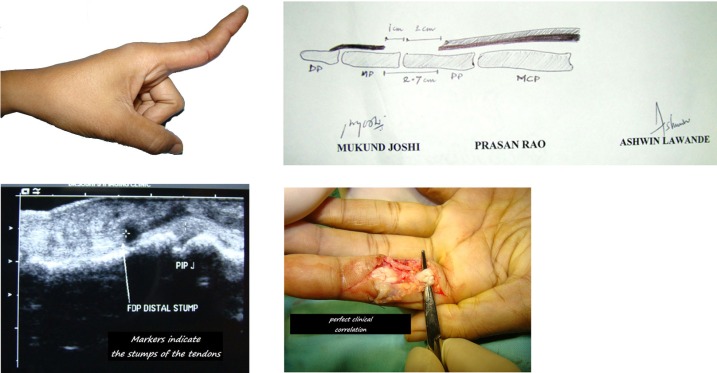
Top left: Flexor Tendon Injury in Zone II. Top right: The corresponding illustration made by the Sonologist, with measurements. Bottom left: The High Frequency Ultrasound image showing the exact location of the tendon stumps. Bottom right: The Operative findings match the illustration and the images perfectly. Picture courtesy: Sudhir Warrier, Mumbai

This is an excellent example submitted by Hemant Kotwal (Ultrasonologist) and Amit Varade (Hand surgeon) from Nasik of how an ultrasound examination reduces the need for extensive exposure while doing a tendon graft. The ultrasonologist has pointed with arrows the two cut ends of the long tendon of the thumb allowing the surgeon to route the tendon graft subcutaneously.

