33. Blowout Fracture of the Orbit
1. Surgical anatomy
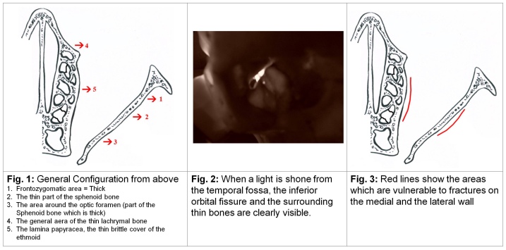
a. The anterior rim of the orbit is somewhat narrower and thicker than the main orbital cavity. The cavity tapers posteriorly and somewhat medially in the form of a cone (Figure 1).
b. The bony structure of the orbit is made up of several bones (see figures below) and is thin in the middle one third particularly the medial, inferior, lateral and superior wall in that order. The floor of the central one third of the orbital cavity is occupied by the inferior orbital fissure and is bound by a very thin part of the maxillary bone medially and the sphenoid laterally (Figure 2). On the orbits medial wall in the central one third lies a very thin bone called the lamina papyracea which in fact is the lateral part of the ethmoid bone (Figure 3). This bone encloses air cells. The whole structure is called the ethmoid sinus. Anterior to the ethmoid bone but not strictly in the central third lies the lacrimal bone which encloses the lacrimal sac. This bone is also quite thin. All in all this arrangement works like a safety mechanism. Should the intra orbital pressure rise suddenly the thin bones give way without affecting the eyeball which is elastic because of its fluid content (aqueous and vitreous humor).
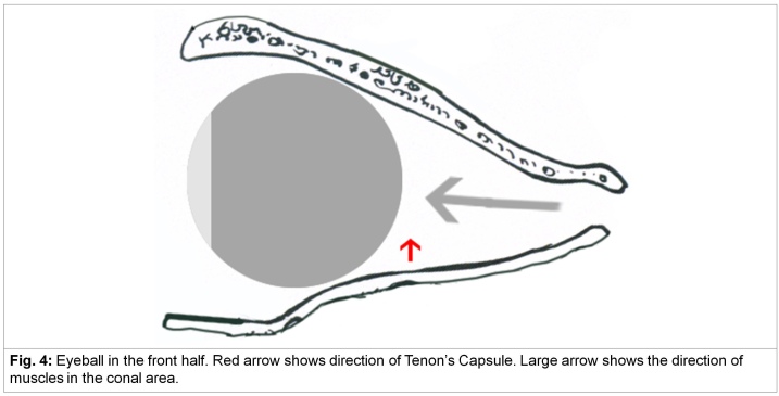
c. The eyeball sits in the front half of the orbit. From behind come the extra ocular muscles which drape the eyeball. This area also contains nerves, vessels, fat and some areolar tissue and is called the cone. In between the eyeball and this cone is a dense fascia called the Tenon’s capsule which together with the orbital septum in front suspends the eyeball. By a rider the extra conal tissue contains periosteum, extra conal fat as enclosed by the orbital septa and muscles such as the levator palpabrae superioris (please see previous chapter 31 on Ptosis) (Figure 4).
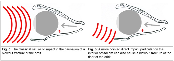
2. The mechanism of the occurrence of a Blowout Fracture: Pan-facial fractures caused by indiscriminate forces which involve the orbit do not follow a set pattern. In fact sometimes in such fractures bones or their parts which form the orbit are pushed within the orbital cavity leading to what is known as a ‘blow in’ fracture. The mechanism of a classical Blowout Fracture involves a forced sudden increase in the normal pressure within the bony orbital cavity leading to fractures allowing the contents of the orbit (both conal and extra-conal to migrate to the extra-orbital space). This migration may or may not be of functional consequence but on the whole in the classical blow out fracture usually saves the eyeball and vision. The functional anatomy of the eyeball is such that the thin part of the bone around the inferior orbital fissure in the orbits floor gives way most frequently. The classic example in modern times occurs in sports injury when an object such as a ball or a stick or a bat hits the protective orbital ring, (the convex part of such an object sometimes hitting the eyeball as well) causing the weaker part of the orbital wall (or walls) to give way. It has been argued that a direct hit to the bone particularly at the junction between the zygoma and the maxilla will cause the weakest part of the bony orbital framework to fracture and may occur in any part of the circumference of the orbits vulnerable central one-third (Figure 5 and 6).
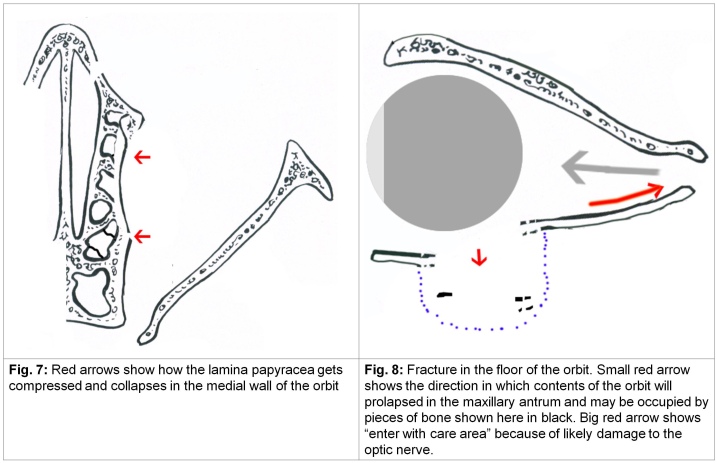
For example, part of the orbital contents might hit medially but rarely enter the space occupied by ethmoidal air cells and only cause a dent (Figure 7) in the lamina papyracea which buckles inwards. This is not true of the maxillary antrum which is already hollow and sits just below the floor of the orbit (Figure 8).
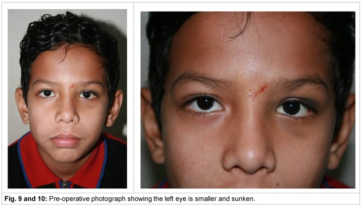
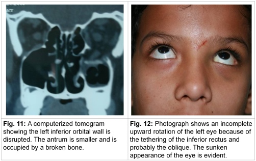
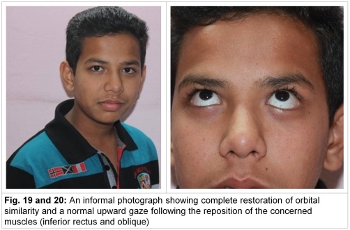
Into this antrum can enter conal tissue such as fat or muscles (inferior oblique or inferior rectus). And this abnormal invagination of muscles might be further complicated by the muscles getting caught in parts of the fractured bone. Both the change of direction of the excursion of the muscle as well as the mechanical hindrance by bony parts can lead to restriction of movements normally effected by these muscles i.e. lower and upper gaze rotation (lower restriction because of change of axis of the muscle, upper restriction because the conal tissue is still stuck in the fracture of the floor – inferior rectus). The inferior oblique when stuck in the fracture will restrict the upper and outward gaze. Also as the orbital space increases the eye ball appears small as compared to the orbit and is usually denoted by the term ‘a sunken eyeball’ irrespective of where the fracture is. The migration of some intra-orbital contents also creates a similar shrinking effect. If the eyeball is greatly displaced or sunk, diplopia can result (Figures 9 & 10).
3. Diagnosis: The principal diagnostic features of a blowout fracture of the orbit are a sunken eye (Figures 9 and 10), restriction of the movement of the eyeball, particularly the downward or upward gaze, echymosis. If the restriction of the movement is severe, and if the eyeballs’ axis is not in consonance with the opposite fellow, will result in diplopia. CT-scans and MRI show the fractures in all its living details and the obliteration of the maxillary sinus (partial or complete) by blood or soft tissue or bony fragments, all help in the diagnosis (see Figure 11 and 12 below in the case report).
4. Treatment: A Blowout Fracture is best not classified because the pathology can range from a severe contusion of the floor of the orbit or a fracture ‘in situ’ without any displacement of tissue to gross fragmentation with abnormal displacement of bone, fat and muscles. The former two may not require treatment though the eye needs to be observed for a post-traumatic/inflammatory tethering. Anti-inflammatory agents might help.
5. In the more severe forms even though diplopia is not present, and the appearance of the eyeball is acceptable, surgical restoration of the broken bone is a treatment of choice to prevent secondary enophthalmos and includes getting the herniated contents from inside the antral cavity into the orbit and then repair or seal the fractured area.
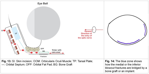
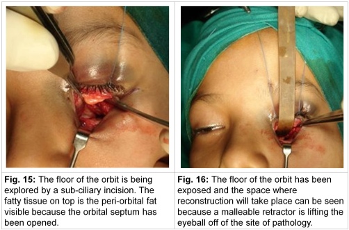
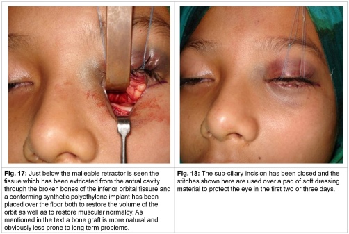
The lamina papyracea which might have buckled in will require to be reduced and more often to be padded up. An intra-ciliary approach is frequently employed. The orbicularis oculi is split below the tarsal plate, the orbital septum which arises from the tarsus and which gets attached to the inferior border of the orbit is opened at this insertion and from here a sub-periosteal dissection is carried out to approach the pathological site. The length from the inferior orbital rim to the optic foramen is about 45 mm and the last posterior 15 mm is an “enter with care zone” for fear that the optic nerve might be injured. This part of the orbit is luckily rarely fractured because it is surrounded by dense bone. Theoretically as well as ideally the defect is best calculated volumetrically as a pre-operative step and must then be filled accordingly. Also ideally an autologous bone graft (calvarial or iliac crest) shaped properly and smoothened towards the ocular surface is placed over the defect (Figures 13, 14, 15, 16, 17 and 18).
The orbit being a tight well occupied space, fixation of the graft or an implant is usually not required. In the case of the fractures of the floor of the orbit all structures which have migrated into the antral cavity are extracted and/or extricated from the fracture site and that this has been successfully done can be gauged by doing “a forced duction test”. This involves holding the sclera with a toothed forceps below the cornea and moving the eyeball in various directions. The wound is closed in layers in which the divided orbital septum is sutured back to the periosteum as the first step. Notice that the incisions in the skin, muscle and the orbital septum and then to the sub-periosteal area of the floor of the orbit are staggered, do not overlap each other and therefore produce a good scar. Late post-operative photographs of the case above are reproduced below (Figure 19, 20).
Note:
1. As in the previous chapters all injuries involving the face and the orbit need a detailed ophthalmic as well as a neurological examination.
2. The line drawings in this chapter are not strictly proportionate to the actual measurements of the eyeball or the orbits.
3. The diagram of the bony orbit is borrowed from Converse’s Plastic Surgery (WB Saunder’s 1990; editor Joseph G. McCarthy) and photoshopped on a computer to explain the text.
4. The compiler of these notes thanks Shailesh Nisal of Nagpur (India) for the almost classical case report of a Blowout Fracture of the Orbit and general help in writing this chapter.
Nitin Mokal, craniofacial surgeon from Mumbai reiterates that a bone graft is always superior to any other alien material and further states that when volumetric replacement for tissues lost following the blowout mechanism is not a consideration, a thin bone graft taken from the anterior surface of the maxillary sinus through an intra-oral approach serves an excellent purpose of blocking of what has been disrupted both in the inferior as well as the medial wall of the orbit. Illustrations reproduced below also show that this procedure can be done by the transconjunctival approach and are self-explanatory.


Leave a comment